|
| The following list of procedures are some of those commonly performed by Mr. Chang. This is not an exhaustive list. Should you require any information about a procedure that is not listed below, please click here to contact main rooms. |
| |
| Upper Urinary Tract Operations |
|
 |
| Bladder Operations |
|
| Prostate Operations |
|
| Testes / Scrotum Operations |
|
|
|
|
|
| |
 |
| |
| Extracorporeal Shock Wave Lithotripsy (ESWL) |
ESWL uses shock waves to break a kidney stone or stones into small fragments that can more easily pass through the urinary tract and out of the body. The non-invasive procedure is performed under general anaesthetic, usually as a day procedure and takes about 30 to 45 minutes. Sometimes, for large or hard stones, more than one treatment may be required before the stone is completely fragmented and able to be passed.
ESWL requires the surgeon to precisely determine the location of the kidney stone(s) with x-ray or ultrasound and then a special machine called a lithotriptor is used to deliver high-energy shockwaves through the patients body to break the kidney stone(s) into small pieces. The kidney stone fragments are usually passed in the urine for a few days following treatment and some patients find that this can cause some temporary mild discomfort.
|
| |
|
|
Back to Top † |
| Laparoscopic Adrenalectomy |
Adrenalectomy is the surgical removal of an adrenal gland, usually for cancer. In most cases this can be performed laparoscopically. The adrenal glands sit on top of the kidneys and secrete hormones into the bloodstream. Click here to learn more about laparoscopy.
|
| |
|
|
Back to Top † |
| Laparoscopic Radical Nephrectomy and Laparoscopic Partial Nephrectomy |
Radical nephrectomy is the complete removal of the kidney for the treatment of kidney cancer. Partial nephrectomy can be considered in any patient with a small (ideally less than four centimetres) localised kidney tumor. Cure rates are similar to radical nephrectomy in these patients.
Laparoscopic radical nephrectomy and laparoscopic partial nephrectomy can be performed through the patient's front (transperitoneal) or through the patient's back (retroperitoneal). The retroperitoneal approach is useful if a patient has had prior abdominal surgery, which could make transperitoneal surgery difficult or impossible.
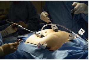
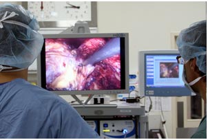
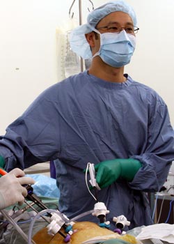
Mr. Chang is experienced in both transperitoneal and retroperitoneal approaches and can tailor an operation to suit a patients circumstances. Mr. Chang also performs laparoscopic partial nephrectomy via the transperitoneal and retroperitoneal approaches. Only a minority of patients will require the traditional open kidney surgery. Click here to learn more about laparoscopy.
|
| |
|
|
Back to Top † |
| Laparoscopic Nephroureterectomy |
| Nephroureterectomy is the removal of the kidney and ureter (the tube connecting the kidney to the bladder). This is most commonly performed for transitional cell carcinoma, a tumour of the lining of the urinary tract. Laparoscopically, this can be performed transperitoneally or retroperitoneally (see section above on laparoscopic radical nephrectomy). Click here to learn more about laparoscopy. |
| |
|
|
Back to Top † |
| Laparoscopic Pyeloplasty |
Pelviureteric junction obstruction (PUJ or UPJ obstruction) is a narrowing at the junction of your kidney with the tube (the ureter), which drains urine from it into your bladder. This narrowing can cause pain, infection or kidney damage. The goal of the pyeloplasty procedure is to remove the narrowed portion of the PUJ and join the two ends back together again. Since the laparoscopic pyeloplasty utilises smaller incisions than the standard open pyeloplasty, the benefits are improved cosmetic result, less pain, and more rapid recovery. Click here to learn more about laparoscopy. |
| |
|
|
Back to Top † |
| Percutaneous Nephrolithotomy (PCNL) |
|
PCNL is performed on patients to remove large or multiple kidney stones. While watching an x-ray screen, the surgeon inserts instruments through the skin and into the kidney via a small incision. The stones are then fragmented and removed under direct vision. At the end of the procedure a tube called a nephrostomy is left in the kidney to allow urine to drain. The nephrostomy tube is removed before the patient is discharged from hospital.
|
| |
|
|
Back to Top † |
| Ureteroscopy |
|
Ureteroscopy is a minimally invasive procedure, which uses an instrument called a ureteroscope to examine the upper urinary tract. The ureteroscope can be rigid or flexible. The ureteroscope is passed through the urethra (the tube that carries urine from the bladder to the outside of the body), into the bladder and then directly into the ureter. The ureter carries urine from the kidneys into the bladder. Ureteroscopy is useful in the diagnosis and treatment of kidney stones or cancers of the upper urinary tract.
|
| |
|
|
Back to Top † |
 |
| |
| Cystoscopy (Rigid or Flexible) |
|
A cystoscopy is a minimally invasive diagnostic medical procedure, which uses an instrument called a cystoscope to look at the inside of the bladder and urethra (the tube that carries urine from the bladder to outside the body). The cystoscope may be a rigid or flexible instrument, which allows for visual inspection of interior surfaces and photography, and may also be used to take biopsies or to remove objects such as stents or stones.
The surgeon inserts the lubricated cystoscope into the urethra and gently advances it into the bladder. Fluid is instilled through the cystoscope to help expand the bladder to create a clear view for the surgeon. Tiny instruments may be inserted through the cystoscope to perform procedures such as collecting tissue samples of the bladder.
Cystoscopy is usually performed as a day procedure under local or general anaesthetic and the procedure itself generally takes between 5 to 10 minutes.
|
| |
|
|
Back to Top † |
| Radical Cystectomy |
|
Cystectomy refers to the surgical removal of all (radical cystectomy) or part (partial cystectomy) of the bladder, usually for the treatment of bladder cancer that has invaded the bladder wall. After the bladder is removed, an ileal conduit may be fashioned using a section of the small intestine, which is then brought out through an opening in the abdomen called a stoma. Urine from the kidneys flows through the ileal conduit, and collects in a bag that attaches on the outside of the body over the stoma, which must be periodically emptied.
An alternative to an ileal conduit is the creation of a neobladder, in which a loop of intestine is surgically fashioned into a pouch. The ureters (which carry urine from the kidneys to the bladder) and the urethra (the tube that normally carries urine from the bladder to outside the body) are attached to the pouch, thereby mimicking the original bladders function. The urine flows from the kidneys into the neobladder, which can be emptied by muscle straining.
|
| |
|
|
Back to Top † |
| Transurethral Resection of Bladder Tumour (TURBT) |
|
TURBT is a surgical procedure, used to diagnose, stage and treat bladder cancer. During TURBT, a rigid cystoscope is passed through the urethra (the tube that drains urine from the bladder to outside the body) to examine the inside of the bladder. An instrument called a resectoscope is used to remove suspicious areas of tissue for biopsy and to burn cancerous cells. Bladder cancer can recur, so TURBT may need to be repeated at intervals to ensure that any new tumours are found at an early stage.
|
| |
|
|
Back to Top † |
 |
| |
| Brachytherapy (HDR or LDR) |
|
Low Dose Rate (LDR) Brachytherapy is a form of radiotherapy used to treat localised prostate cancer. The procedure involves the permanent implantation of small radioactive seeds directly into the prostate using a needle, which is inserted through the perineum (the area between the genitals and the anus) under ultrasound guidance. The seeds emit a dose of radiation that affects the tissue in a small area with the aim of killing the cancer cells.
High Dose Rate (HDR) Brachytherapy involves placing hollow rods into the prostate via the perineum and the radiation dose is delivered through the rods over a few days. Patients considering Brachytherapy will also be referred to a Radiation Oncologist for a specialised opinion about their suitability for the treatment.
|
| |
|
|
Back to Top † |
| Laparoscopic Radical Prostatectomy |
|
Radical Prostatectomy is a surgical procedure in which the entire prostate and the seminal vesicles (glands that produce ejaculatatory fluid) are removed. Radical prostatectomy is indicated for patients with prostate cancer that is confined to the prostate gland. Radical prostatectomy requires that the entire prostate be removed to ensure that no cancer cells remain. This is because the cancer cells may be distributed throughout the prostate gland in more than one area.
Laparoscopic Radical Prostatectomy is a minimally invasive surgical technique that uses small incisions rather than one large surgical incision to remove the prostate. Click here to learn more about laparoscopy.
|
| |
|
|
Back to Top † |
| Open (Millins) Prostatectomy |
Open Prostatectomy is a surgical procedure not commonly used to treat Benign Prostatic Hyperplasia (BPH). This surgery is usually reserved for patients with extremely large prostates, or in patients where Trans Urethral Resection of the Prostate (TURP) is not possible. See section below on TURP.
During the procedure the surgeon makes an incision in the lower abdomen. The inner portion of the prostate is shelled out or enucleated. Like TURP, the procedure also involves the temporary placement of a urinary catheter. The hospital stay for an open prostatectomy is slightly longer than for TURP, typically around 3-4 days.
|
| |
|
|
Back to Top † |
| Trans Urethral Resection of the Prostate (TURP) |
|
Transurethral resection of the prostate (TURP) is a surgical procedure used to treat bladder outlet obstruction due to Benign Prostatic Hyperplasia (BPH), more commonly known as prostate gland enlargement.
The prostate gland sits below the bladder, surrounding the urethra (the tube that carries urine from the bladder to outside the body) and is responsible for prostatic fluid production for semen. When the prostate becomes enlarged, it puts pressure on the urethra, blocking the flow of urine, causing voiding difficulties which may include symptoms such as difficulty starting urination, a poor urinary stream, dribbling at the end of urinating, straining or stopping/starting while urinating, an urgent or frequent need to urinate, feeling of incomplete emptying after urinating, blood in the urine or urinary tract infection. Treatment of prostate gland enlargement depends on an individuals signs and symptoms. Treatment may be medical (i.e. medication) or surgical (e.g. TURP).
After the spinal or general anesthetic takes effect, the surgeon passes an instrument called a resectoscope into the urethra (the tube that carries urine from the bladder to outside the body). The resectoscope contains an electrical loop that cuts or vaporises tissue and seals blood vessels. Using the loop, the surgeon removes the obstructing tissue from the inside of the prostate, creating a cavity. Only the interior part of the prostate is removed. The resected tissue is then flushed out of the bladder at the end of the operation. A flexible tube called a urinary catheter is passed into the bladder via the urethra and drains urine into a bag. The urinary catheter is generally removed the next day.
TURP generally requires overnight hospitalisation. The operation typically takes between 30 to 60 minutes. Mr. Chang usually performs a bipolar TURP (Gyrus), which allows the resection to be performed using a saline irrigation, and reduces the complications associated with traditional monopolar TURP.
|
| |
|
|
Back to Top † |
 |
| |
| Orchiectomy |
|
Orchiectomy refers to the surgical removal of a testicle. The procedure is commonly performed where a testicular cancer is suspected. Other tests such as a scrotal ultrasound and/or tumour marker blood test may assist with diagnosis and aid management.
Suspicious tumours are treated by orchiectomy through a small groin incision. Sometimes a testicular prosthesis may be inserted at the time for cosmetic reasons.
If cancer is confirmed further treatment may include chemotherapy, radiotherapy or surgical removal of lymph nodes in the abdomen.
|
| |
|
|
Back to Top † |
| Orchidopexy |
|
Orchidopexy is a surgical procedure where an undescended testicle is brought down to the scrotal sac and secured in place using stitches. The procedure is commonly performed in infants and adolescents. Untreated, undescended testes are associated with an increased risk of testicular cancer in men.
Orchidopexy is most often performed to treat testicular torsion, which is a urological emergency. Testicular torsion is the twisting of a testicle on its connection, which if left untreated, may result in the testicle dying due to lack of blood supply. The surgeon makes a small incision in the scrotum and untwists the testicle. If the testicle has died, it is removed. If it appears to be healthy, the testicle is stitched in place to prevent it from re-twisting and the incision is closed. The surgeon will usually stitch the unaffected testicle in place to prevent torsion on that side as a preventative measure.
|
| |
|
|
Back to Top † |
| Bilateral Vasectomy |
|
A vasectomy is a minor surgical procedure designed to interrupt the sperm transportation system between the testicle and the urethra by blocking the vasa deferentia.
A small incision is made in the scrotum so the vas deferens on each side can gently be lifted out, cut, then tied or cauterized and put back in place.
|
| |
|
|
Back to Top † |
| Vasectomy Reversal |
|
Vasectomy reversal restores fertility by using microsurgery to reconnect the ends of the severed vas deferens, which is located in each side of the scrotum.
|
| |
|
|
Back to Top † |
| |